

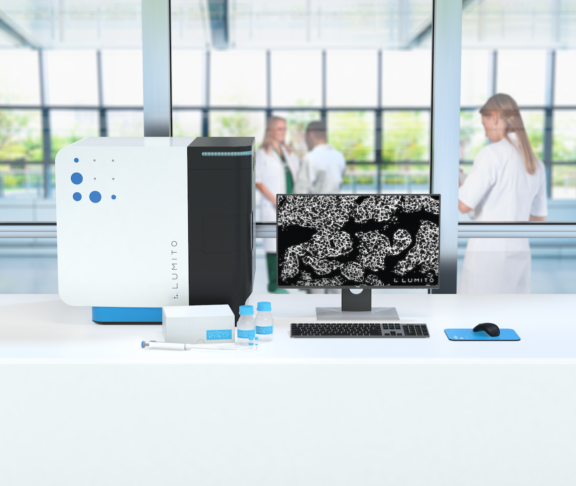
Lumito offers the biomedical research community the next generation of IHC detection systems. Using Lumito’s digital images, produced by a new imaging technique, histology researchers have a more complete basis for analysis and clinical diagnosis.
The product, Scizys by Lumito, includes a digital scanner and a reagent kit with UCNPs (upconversion nano particles). The characteristics of the particles allow the scanner to produce dual modality images. For each scanned sample, two images are acquired. One for morphological information including chromogenic IHC and one for IHC with UCNP as reporter. The images are completely separated into two channels, but fully coordinated and stackable. This allows the users to study all information in the same section.
In the future, the platform may provide additional multiplexing channels.
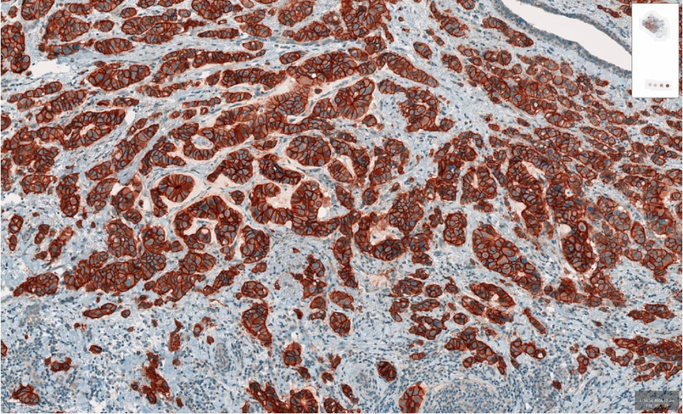
Brightfield chromogen channel. HER2 with DAB.
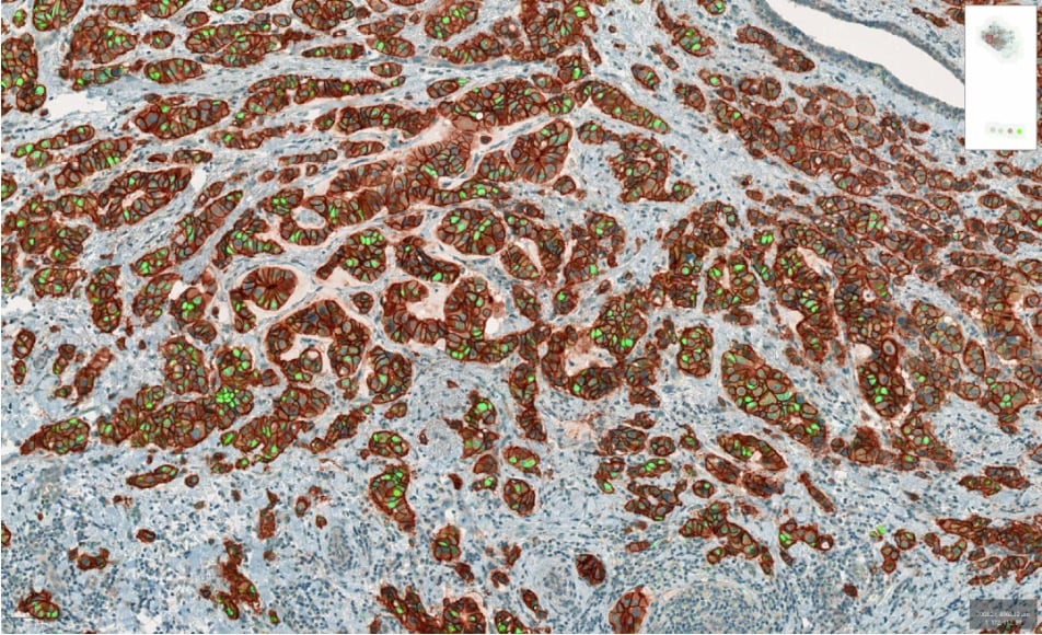
UCNP. With Lumito’s UCNPs, a higher degree of plexing for IHC samples can be explored, avoiding the need having to perform a new staining.
Requires neither frozen tissue nor darkroom
The images have a higher contrast and granularity compared to traditional immunohistochemistry, which is likely to better enable decision support tools based on image analysis and machine learning (AI). In addition, this unique technology is completely free of autofluorescence and has a very low detection threshold. The ambition is to launch in the first part of 2023 in the research labs segment.
Preliminary studies to identify the most significant benefits
By collaborating with research groups at the forefront of their respective research fields, knowledge is gained on where Lumito’s technology and solution works best, provides the most benefit and where it meets the needs of researchers. Valuable knowledge that benefits further development of the clinical product.
Good results in completed pre-study with Umeå University
The pre-study was conducted in collaboration with Umeå University and a research group led by Associate Senior Lecturer Daniel Öhlund. The research group aimed to identify how Lumito’s UCNP technique may be used to improve the ability to visualize protein expression in pancreatic cancer. A scientific paper, ”Extracellular galectin 4 drives immune evasion and promotes T-cell apoptosis in pancreatic cancer”, is now published in the scientific journal Cancer Immunology Research.
– Using Lumito’s imaging technique, we have investigated, among other things, whether a certain protein spreads via secretion from the cancer cells into the tumor’s supporting tissue, the tumor stroma. Lumito’s technique has provided better possibilities, compared to other immunohistochemical methods, to visualize the penetration of secreted proteins into the tumor stroma, says Daniel Öhlund.
The scientific paper’s first author is Tommy Lidström, Ph.D., Postdoc.
– In comparison to DAB staining, Lumito’s technique is more suited for signal quantification since the brown DAB staining signal becomes saturated very quickly, making quantification more difficult, says Tommy Lidström.
Current pre-studies
Lumito’s first international pilot study
A pilot study, aimed at investigating the potential of Lumito’s UCNP to detect the deposition of immune complexes and complements in renal biopsies, has been initiated in collaboration with Dr Kishore Gopalakrishnan’s research group at University Hospitals Coventry and Warwickshire NHS Trust in the UK.
Dalarnas forskningslaboratorium
Under the leadership of Laboratory Manager Helena Hermelin at the Dalarna Research Laboratory, a pilot study has been initiated to analyze whether Lumito’s UCNP is compatible with current analytical methods for breast cancer.
– The advantage of UCNP is that in the same hematoxylin-stained tissue section, in addition to HER2, it is possible to stain multiple biomarkers simultaneously with the ability to switch between the different images to determine the presence of different marker epitopes, comments Helena Hermelin.
Uppsala University
A research group at Uppsala University and the Department of Immunology, Genetics and Pathology is conducting a pilot study to determine whether Lumito’s technique can provide better quantification and contribute to a more reliable assessment than the current limited and highly subjective analysis methods used for clinical diagnostics. UCNP may allow a more reliable assessment of whether the tumor can be treated with PD-L1 inhibitors.
Are you interested in getting in touch?
If you are interested in getting in touch with Lumito for further information, please contact Mattias Lundin, CEO, e-mail: [email protected]. You are also welcomed to visit the website www.lumito.se/en.