Lumito presents a new innovative digital imaging technology. With the help of the company’s digital images, pathologists and researchers can obtain a more comprehensive histology-based foundation for their analyzes and clinical diagnostics.
– The ambition is to offer a powerful tool and meet fast and safe tissue diagnostics requirements. The plan is to launch our first product, SCIZYS by Lumito, for research laboratories at the end of 2022 and then, according to the plan, a clinical product, says Andreas Johansson, technical director at Lumito.
A WSI scanner, combined with Lumitos UCNP reagents (Up Converted Nano Particles), provides high-quality IHC imaging with unique qualities. The scanner also takes images of tissue samples traditionally stained with hematoxylin and chromogenic immunohistochemistry (IHC).


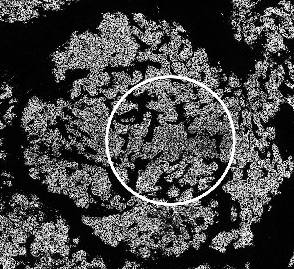
The images show cell nuclei and HER2 in identical cells in the same tissue section.
– The image on the left shows a tissue section with traditional histological staining that illustrates the presence of all cells (hematoxylin staining with blue nuclei) in a tissue section.
– The image on the right shows the same tissue section with the cancer cells, labeled specifically with Lumitos UCNP (white, Her2 grade 3 positive cancer cells).
Higher analysis quality and shorter analysis time
The technology enables obtaining images with higher contrast, increased dynamic range, and reduced non-specific background. Through higher analysis quality and shorter analysis time, the diagnostics of tissue samples can be significantly improved.
– It is also possible to switch between visualizing the morphology or our unique UCNP IHC labeling. The system’s flexibility also allows visualization of both readings simultaneously, superimposed in the same image. This will provide more detailed information, but without issues with overlap and non-specific labeling that traditional technology provides, says Andreas Johansson.
Lumito has recently completed a preliminary study with Umeå University under the leadership of assistant university lecturer Daniel Öhlund, with positive results. The research group has mapped how the company’s UCNP technology can be used to improve the ability to visualize protein expression in pancreatic cancer.
– Using Lumito’s imaging technology, we have, among other things, investigated whether a particular protein spreads via secretion from the cancer cells to the tumor’s supporting tissue, the tumor stroma. Lumito’s technology has brought better opportunities, compared to other immunohistochemical methods, to illustrate the penetration of secreted proteins into the tumor stroma, says Daniel Öhlund.
– We have several studies underway but are always interested in getting in touch with more research groups to map out where our product brings the most value, concludes Andreas Johansson.
Want to learn more? Please contact Product Specialist Tim Hofvendahl, [email protected].


